
FIGURE 11.
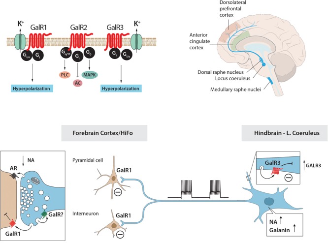
The galanin–locus coeruleus (LC) system in stress and depression: A hypothesis. The hypothesis is built on animal (rat) experiments showing that (i) galanin and GalR1 (top left) are present in LC NA neurons; (ii) galanin mRNA levels are increased during stress; (iii) galanin via GalR1 autoreceptors inhibits firing of LC neurons; and (iv) indirect evidence that galanin can be released from soma-dendrites of LC neurons. The second corner stone is results from two studies on human postmortem brain with ISH and qPCR. Five regions from postmortem brains from depressed subjects who committed suicide and controls were studied and are shown, including LC that projects to anterior cingulate and dorsolateral prefrontal cortices (top right). The results show that also in humans (i) the NA LC neurons express in any case galanin and GalR3 (top left). GalR1 and GalR3 probably have similar transduction mechanisms (top left). Under ‘normal’ firing only noradrenaline is released in forebrain. A situation after severe stress is depicted in the lower panel: LC neurons burst fire (lower panel, middle), NA and galanin are released from nerve endings in cortex (lower panel, left) and dendrites in the LC (lower panel, right), the latter in an attempt to prevent overexcitation (a resilience mechanism). To replace released peptide, galanin transcript levels and synthesis increase, and also GalR3 is upregulated (lower panel, right). The increased release, together with elevated galanin and GalR3 levels, result in a too strong inhibition and decreased NA levels in the forebrain (maladaptation) (lower panel, left), possibly contributing to depressive symptoms. HiFo, hippocampal formation. Drawing by Mattias Karlén.