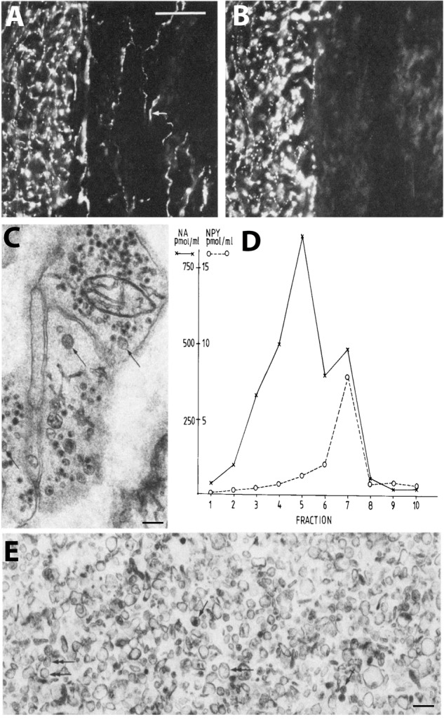
FIGURE 2.

Coexistence and subcellular distribution of neuropeptide Y (NPY) and noradrenaline (NA) in the rat vas deferens. (A,B) Immunohistochemical visualization of NPY- (A) and tyrosine hydroxylase (TH)-(B) positive nerve terminals in adjacent sections. Overlapping, dense NPY and noradrenergic networks are seen in the muscle layer. Note sparse NPY-only positive nerves (arrow) in the subepithelial region, possibly cholinergic nerves. (C) Electron microscopic micrograph of several nerve terminal profiles in the muscle layer after potassium permanganate (KMnO4) fixation, showing small synaptic vesicles with a dense core and LDCVs. The dense core indicates presence of NA both in the synaptic and LDCVs (cf. Figure 1D). No profiles without small vesicle with a dense core are seen, suggesting a pure adrenergic innervation of the muscle layer. (D,E) Subcellular distribution of NA (x) and NPY (o) in a density gradient of rat vas deferens. There is only one peak for NPY (fraction 7; E), whereas there are two peaks for NA (fraction 5 and 7), tentatively representing synaptic vesicles and LDCVs, respectively. Note many LDCVs (arrows), as well as many vesicles of the same size but without dense core (double-headed arrow). The peptide is only present in the heavy fraction (in agreement with Figures 1C,E), whereas NA is present also in the light one (in agreement with Figure 1D). On the abscissa, totally recovered sedimentable substance is given as picomoles per milliliter after centrifugation at 145,000 × gmax for 45 min. On the ordinate, density gradient fractions 1–10 are given, corresponding to the following sucrose molarities: 1 (0.26 M), 2 (0.32 M), 3 (0.47 M), 4 (0.56 M), 5 (0.69 M), 6 (0.74 M), 7 (0.84 M), 8 (0.91 M), 9 (0.98 M), 10 (1.2 M). Recoveries of NA = 70%, of NPY = 65%, and of protein = 87%. Reprinted from Fried et al. (1985), with permission.
