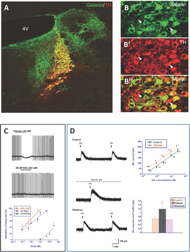
FIGURE 5.

(A–B”) Immunofluorescence micrographs of the dorsal pontine periventricular region of mouse after double-staining of a section with antibodies to galanin (green) and tyrosine hydroxylase (TH) (red), the rate-limiting enzyme for catecholamine synthesis and thus a marker for NA neurons. Note that both antibodies stain neurons in the locus coerulus (LC) (B,B’), whereby many (yellow, B”), but not all TH-positive neurons express galanin [arrowheads point to TH-only neurons (red), apparently lacking galanin] (B’). Galanin is also present in many structures outside the LC. Colchicine treated animal. Courtesy Joanne Bakker and Mingdong Zhang. Bar for (A) 200 μm, for (B–B”) 20 μm. (C) Effect of galanin and the GalR2 agonist AR-M1896 on LC neurons (upper two traces), and the dose–response curves of galanin (red), the AR-M1896 (green) and the mixed GalR1-GalR2 M961 agonist (magenta) (lower trace). Note strong hyperpolarization of galanin and a less strong effect of M961, whereas that AR-M1896 hardly causes any effect at all. From Ma et al. (2001). (D, left panel) Effect of galanin on the response of LC neurons to NA. NA (applied from a pipette at the arrowhead) induces a persistent outward current (upper trace). When galanin (0.1 nM) is present, the NA-induced outward current is enhanced, and the duration is prolonged (middle trace). After wash out of galanin, the amplitude and duration of the NA response was similar to that seen before galanin administration (lower trace). (D, right panel) Effect of galanin on dose-response (upper figure) and duration (lower figure) of NA. The NA dose-response curve is shifted to the left, when galanin (0.1 nM) is present (upper figure). The duration of the NA-induced current is increased in the presence of galanin (lower figure). ∗∗P < 0.01. From Xu et al. (2001) with permission.
