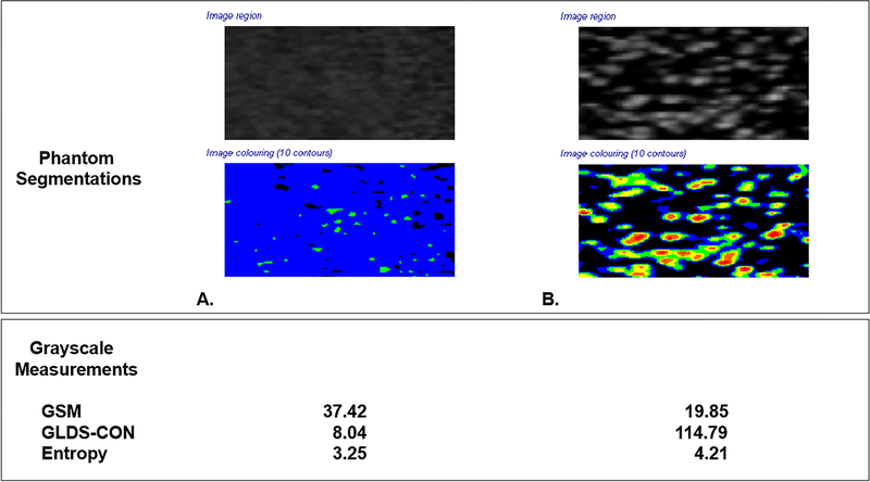
Figure 2. Ultrasound Phantom Images.

Grayscale segmentations from two different ultrasound imaging phantoms with different acoustic scattering properties. Phantom Images were obtained with the Acuson S2000 ultrasound system and 9L4 transducer (Siemens Medical Solutions, Malvern, PA). All instrumentation settings remained constant for both phantom images.
Panel A. Ultrasound phantom images (404GS precision small parts grayscale phantom, Gammex, Middleton, WI) with tissue-mimicking material at speed of sound 1540±10 m/s and attenuation coefficient 0.7±0.01 dB/cm-MHz showing scatterer properties sufficient to yield a fully developed speckle pattern which demonstrates higher GSM, lower GLDS-CON and lower entropy values.
Panel B. Ultrasound phantom images with 800 glass bead scatterers/cm3 demonstrating lower GSM, higher GLDS-CON, and higher entropy values. Note variation in grayscale appearance and corresponding colorized values in the segmented images.
Colorized segmentations correspond to the following grayscale values: 0–25 = black, 26–50=blue, 51–75=green, 76–100=yellow, 101–125=orange and 126–255=red
B and X in an image represent extra arterial landmarks used for image reproducibility.
GSM = Grayscale median
GLDS-CON = Gray level difference statistic – contrast
