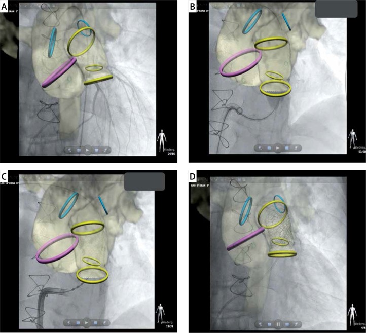
Figure 3.
Live three-dimensional guidance of percutaneous pulmonary valve implantation. Magnetic resonance imaging derived three-dimensional roadmap (see Figure 2) was utilized to guide successive steps of the intervention: selective coronary artery angiography (A), pre-stenting with implantation of two covered stents (B), placement of a 26 mm Sapien 3 valve (Edwards) (C) and final angiography (D)

