Fig. 3.
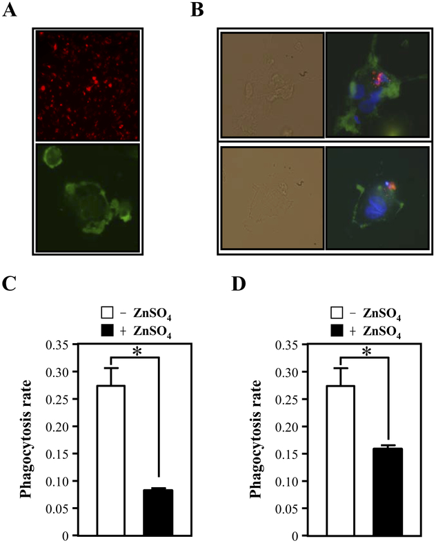
Conditioned medium from U373MG cells overexpressing CEBPD inhibits the phagocytosis of apoptotic SH-SY5Y cells or primary neurons by macrophages. (A) SH-SY5Y cells were labeled with PKH26 red fluorescent membrane tracer (upper panel). THP-1 macrophages were stained with F4/80 (green) (bottom panel). (B) The SH-SY5Y cells (labeled with PKH26 red fluorescence) were induced to undergo apoptosis by irradiation and were then incubated with activated macrophages (labeled with F4/80 green fluorescence). The upper panel shows an apoptotic cell phagocytosed by a macrophage. The lower panel shows an apoptotic cell attached to the cell membrane of a macrophage. The bright field panel represents the phase contrast image. (C) The apoptotic SH-SY5Y cells or (D) apoptotic primary neurons were added to adherent monocyte-derived macrophages incubated with control (−ZnSO4) or CEBPD overexpressing (+ZnSO4) conditioned media for 30 minutes. Phagocytosis was quantified by flow cytometric analysis of fluorescent particles. The phagocytosis ratio was calculated by dividing the number of stained cells by the total number of macrophages (mean ± SD, n = 3, * p < 0.05 by Student t test).

