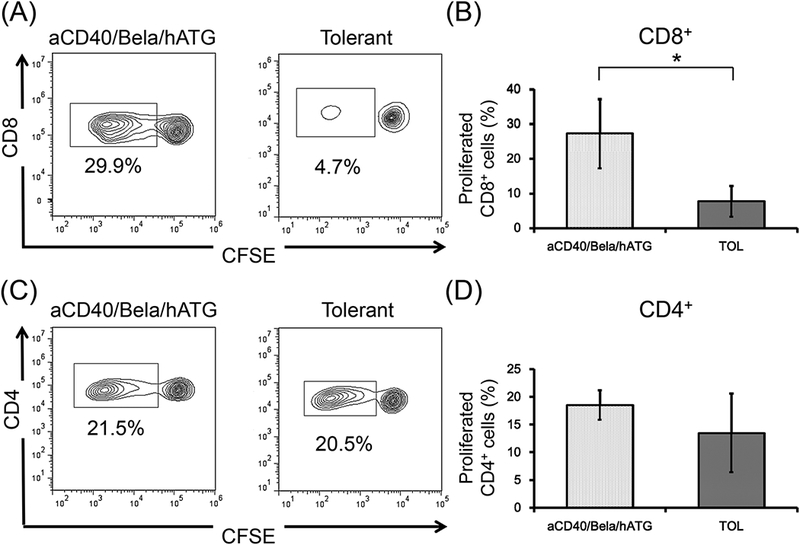
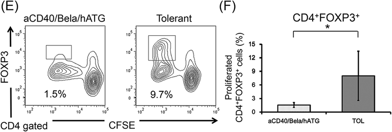
Figure 5: Neither loss of anti-donor CD8+ responses nor donor-antigen-specific Treg expansion was observed in aCD40/Bela/hATG treated animals in vitro.


CD3+ T cells isolated from aCD40/Bela/hATG treated recipients were labeled CFSE and were cultured with irradiated self or donor peripheral mononuclear cells (PBMCs) for 5 days. Cultured cells were then stained for CD4, CD8, and FOXP3. Representative flow cytometric data (A, CD8; C, CD4; E, CD4 gated FOXP3) and mean % proliferation relative to the response to self antigens (B, CD8; D, CD4; F, CD4 gated FOXP3) are shown compared with those of previously published results in 8 tolerant recipients.14 The loss of anti-donor CD8+ T cell response was not observed in the aCD40/Bela/hATG treated recipients, which was significantly higher than those observed in the tolerant recipients (A and B). Substantial anti-donor CD4+ T cell proliferation was observed in the aCD40/Bela/hATG treated animals, comparable to that observed in tolerant recipients (C and D). Among these proliferated CD4+ cells after donor stimulation, a significant proportion of proliferating CD4+ cells were found in FOXP3 intolerant recipients, while such FOXP3+ cell proliferation was minimal in the aCD40/Bela/hATG treated animals (E and F). Proliferated cells (%) = Proliferated cells (%) with donor antigens - Proliferated cells (%) with self antigens. Data are presented as the mean ± SD. *P<0.05.
