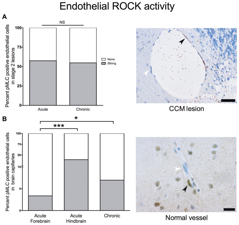
Figure 3. Endothelial Rho kinase protein (ROCK) activity in Stage 2 lesions and brain capillaries.
(A) (Left) No significant difference was observed between the ROCK activity in endothelial cells of acute Cdh5iCreERT2Krit1ECKO (n=93 cells counted in 5 lesions from 3 mice) and chronic Krit1+/−Msh2−/− lesions (n=358 cells counted in 13 lesions from 3 mice). (Right) Representative image of a Stage 2 lesion stained for ROCK activity showing an endothelial cell with strong ROCK activity (brown staining; black arrowhead) and another with no ROCK activation (blue staining; white arrowhead). Scale bar is 50 μm. (B) (Left) Krit1+/−Msh2−/− brain normal capillaries (n=583 cells from 3 mice) had a significantly higher (p=0.01) strong ROCK activity compared to Cdh5iCreERT2Krit1ECKO normal capillaries (n=445 cells from 3 mice) far from lesions in the forebrain. Endothelial cells of Cdh5iCreERT2Krit1ECKO (n=179 cells from 3 mice) normal capillaries near lesions in the hindbrain had higher strong activity (p<0.001) than those far (n=445 cells from 3 mice) from lesions in the forebrain. (Right) Representative image of a normal capillary stained for ROCK activity. Scale bar is 20 μm. All p values were considered to be statistically significant at **p<0.01 or ***p<0.001.

