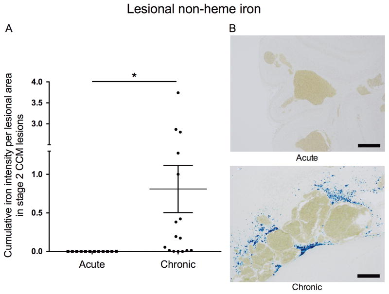
Figure 4. Non-heme iron deposition in Stage 2 CCM lesions in acute and chronic models.
(A) Lesions from chronic Krit1+/−Msh2−/− (16 lesions from 13 mice) harbor significantly higher (p=0.03) integrated density of non-heme iron deposition per lesional area compared to lesions from acute Cdh5iCreERT2Krit1ECKO (12 lesions from 9 mice). (B) Representative non-heme iron deposition (Perls blue stain) in Stage 2 lesions from an acute Cdh5iCreERT2Krit1ECKO model (upper) and a chronic Cdh5iCreERT2Krit1ECKO model (lower). Scale bars are 200 μm. All p values were considered to be statistically significant at *p<0.05.

