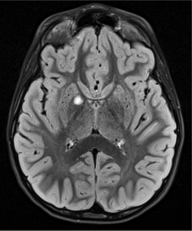
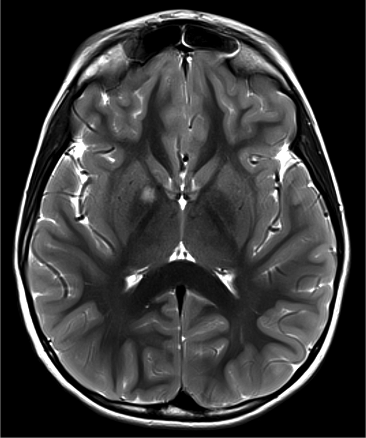
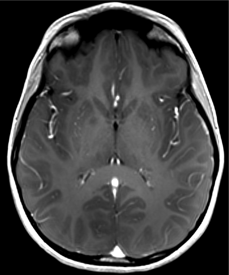
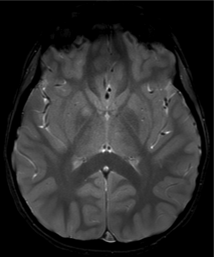
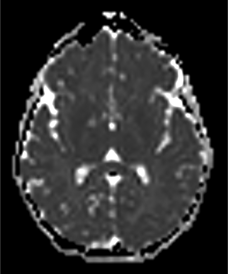
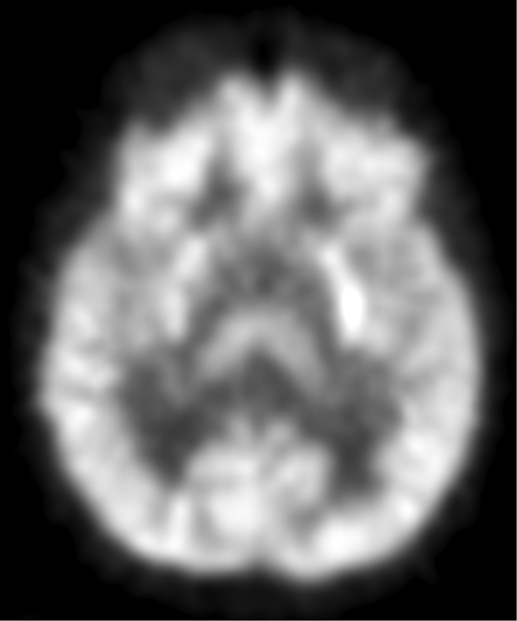
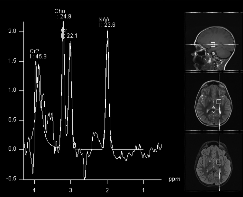
Figure 1.
Imaging characterization of the brain lesions. Except for the location, the imaging characteristics of the lesions were similar for all 3 patients. These images are from Individual 3 at age 8 years 1 month (except for the PET scan, which was obtained at age 7 years 8 months). The FLAIR (A) and T2-weighted (B) images demonstrate a hyperintense lesion with infiltrative qualities in the anterior part of the right globus pallidus. The lesion is hypointense and does not enhance on post-contrast T1-weighted images (C). Susceptibility-weighted images (D) do not demonstrate metal or mineral deposition within the lesion, and the ADC map (E) derived from diffusion tensor imaging (DTI) demonstrates elevated diffusivity within the lesion. FDG-PET(F) demonstrates that tracer uptake within the lesion is indistinguishable from surrounding tissues. A long-echo single voxel 1H MR spectrum (PRESS technique, TE=144 ms, voxel volume = 1 cm3) of the lesion (G) does not differ significantly from a spectrum obtained from the contralateral normal side (H) in the same individual. Maps of relative cerebral blood volume (rCBV), relative cerebral blood flow (rCBF), and relative mean transit time (rMTT) derived from dynamic susceptibility contrast perfusion imaging were unremarkable (images not shown). On all of the imaging sequences, the lesions were homogeneous, without discernable internal structure.

