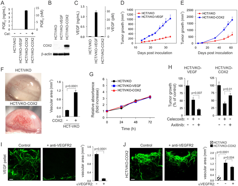
Figure 3. Expression of VEGF or COX-2 in HCT/VKO cells promotes tumorigenesis.
(A) PGE2 concentration in the conditioned media of HCT/VKO-derived cell lines as measured by ELISA. PGE2 was below the detection limit in the HCT/VKO, HCT/VKOVEGF, and celecoxib-treated HCT/VKO-COX2 cell lines.
(B) COX-2 protein in HCT/VKO-derived cell lines as measured by Immunoblotting. β-actin was used as a loading control.
(C) VEGF concentration in the conditioned media of HCT/VKO-derived cell lines as measured by ELISA. VEGF was not detected in the HCT/VKO and HCT/VKO-COX2 cell lines.
(D) Tumor growth rates of HCT/VKO and HCT/VKO-VEGF subcutaneous xenografts.(E) Tumor growth rates of HCT/VKO and HCT/VKO-COX2 subcutaneous xenografts.
(F) Tumor spheroids were implanted into the cornea to evaluate the role of COX-2 in tumor angiogenesis. Right panel: Quantification of corneal angiogenesis. P < 0.0001.
(G) An Alamar blue assay was used to measure the relative growth of HCT/VKO-derived cell lines in vitro.
(H) The response of HCT/VKO-VEGF (left panel) or HCT/VKO-COX2 (right panel) subcutaneous xenografts to celecoxib and axitinib was evaluated in vivo.
(I) The corneal assay was used to measure the effect of systemic DC101 anti-VEGFR2 antibody treatment on VEGF-induced vascular sprouting in vivo. Bar: 100 μm.
(J) The corneal assay was used to measure the effect of systemic DC101 anti-VEGFR2 antibody treatment on angiogenesis induced by HCT/VKO-COX2 spheroids. Bar: 100 μm.
Data are presented as mean ± SD (A, C, and G) or mean ± SEM (D, E, F, H-J).

