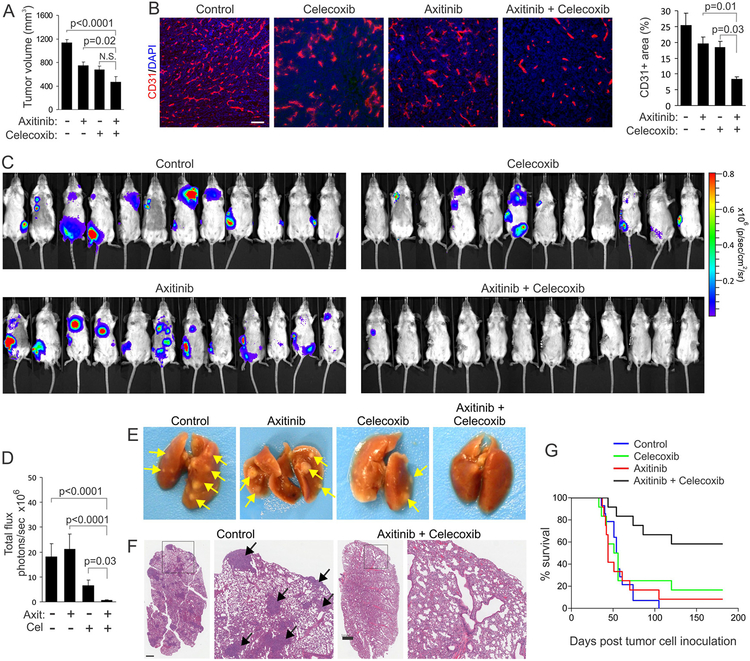
Figure 6. Dual celecoxib/axitinib therapy blocks spontaneous metastasis and extends survival.
(A) The orthotopic growth of 4T1-luc breast tumors in the mammary fat pad was evaluated after treatment with axitinib and celecoxib.
(B) CD31 immunofluorescent staining (red) was used to assess blood vessel densities in 4T1-luc primary tumors after treatment with axitinib and celecoxib. Bar: 100 μm. Right panel: quantification of CD31 vessel staining.
(C) Bioluminescent imaging of metastasis 35 days after injection of 4T1-luc cells into the mammary fat pad. Primary tumors were resected when they reached a size of ~1000 mm3.
(D) Quantification of tumor burden shown in (C) applying a maximum luminescence threshold of 3×10⁷. N.S.: non-significant (n=12/group).
(E) Macroscopic appearance of 4T1-luc tumor burden in the lung. Arrows: tumor nodules.
(F) Microscopic appearance of 4T1-luc tumor burden in the lung after H&E staining. Arrows: tumor nodules. Bar =1 mm.
(G) Kaplan-Meier survival curves were used to asses the impact of celecoxib and axitinib treatment on overall survival of the mice imaged in (C) and (D). Treatments began one day after tumor cell inoculation and continued for the duration of the study. Log-rank analysis: p=0.0001 combination vs. control, p=0.02 combination vs. celecoxib, p=0.001 combination vs. axitinib. All other comparisons were non-significant.
Data are presented as mean ± SEM.

