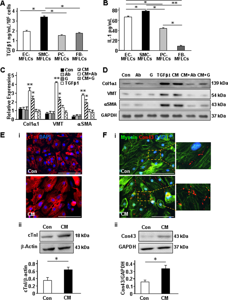
Figure 3. Conditioned medium from cultured hiPSC-SMC–myofibroblast-like cells (MFLCs) alters protein expression in cultured hiPSC-NMCCs and cardiomyocytes.
(A-B) The media of cultured hiPSC-EC–, -SMC–, -pericyte (PC)–, and -fibroblast (FB)–MFLCs was collected, and (A) TGFβ1 and (B) IL-1 levels were evaluated via ELISA. (C-D) A combined population of hiPSC-ECs, -SMCs, -pericytes, and -fibroblasts was cultured with TGFβ1, with an anti-TGFβ antibody (Ab), with the TGFβ-receptor 1-blocker galunisertib (G), with conditioned medium from hiPSC-SMC–MFLCs (CM), with CM and an anti-TGFβ antibody (CM+Ab), with CM and galunisertib (CM+G), or under standard conditions (Con); then, Col1α1, VMT, and αSMA (C) mRNA and (D) protein levels in the medium from the cultured cells were evaluated via quantitative RT-PCR and Western blot, respectively. (E-F) hiPSC-derived cardiomyocytes were cultured with CM from hiPSC-SMC–MFLCs or under standard conditions; then, (E) Cardiac troponin I (cTnI) and (F) connexin 43 (Con43) expression were evaluated via (i) immunofluorescence and (ii) Western blot. Nuclei were counterstained with DAPI, and Western blots of β-actin or GAPDH levels were evaluated to confirm equal loading. Bar=100 μm; *P<0.05, **P<0.01 versus Control, CM+AB, and CM+G; One-way ANOVA followed by Tukey post-hoc test for A, B, C; Two-tailed Student’s t-test for E, F. n=3–4 independent experiments.

