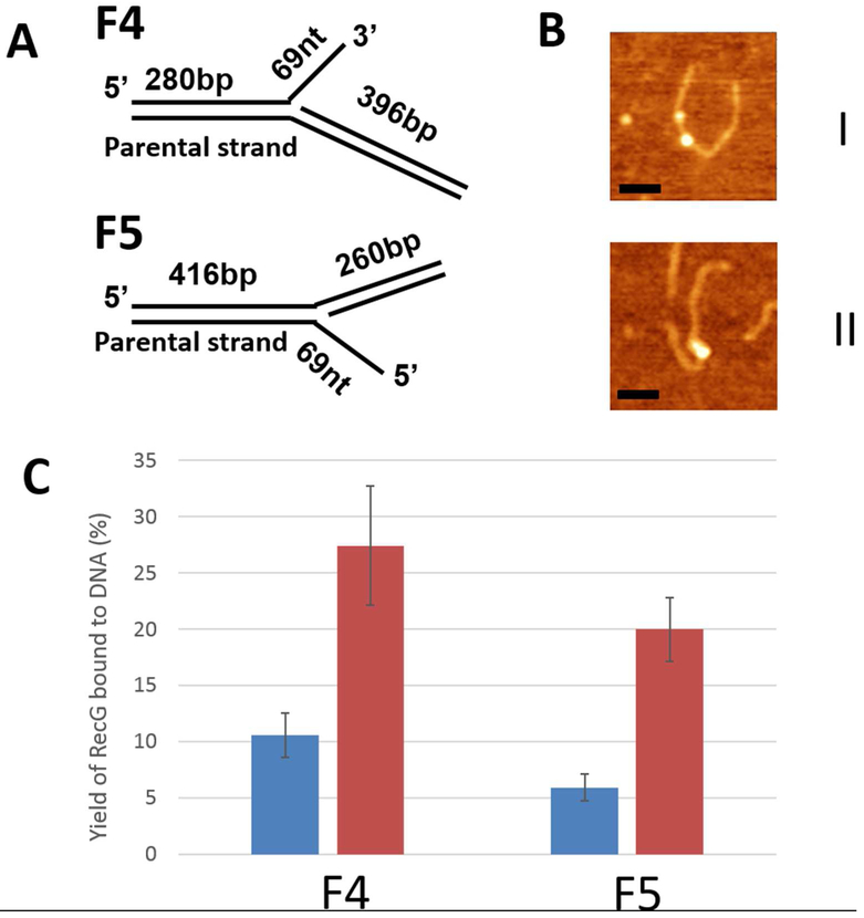
Figure 1.
The interaction of RecG with stalled replication fork substrates. (A) Replication fork substrate F4 consists of two DNA duplexes (280 bp and 396 bp) with a ssDNA arm of 69 nt. The F5 construct has duplex segments of 260 bp and 416 bp, and a 69 nt 5′ ssDNA. (B) Typical AFM images of SSB-RecG complexes with F4 (I) and F5 (II). Bars are 50 nm. (C) Yields of SSB-RecG complexes with replication fork substrates calculated in the absence (blue bars) and presence of SSB (red bars).

