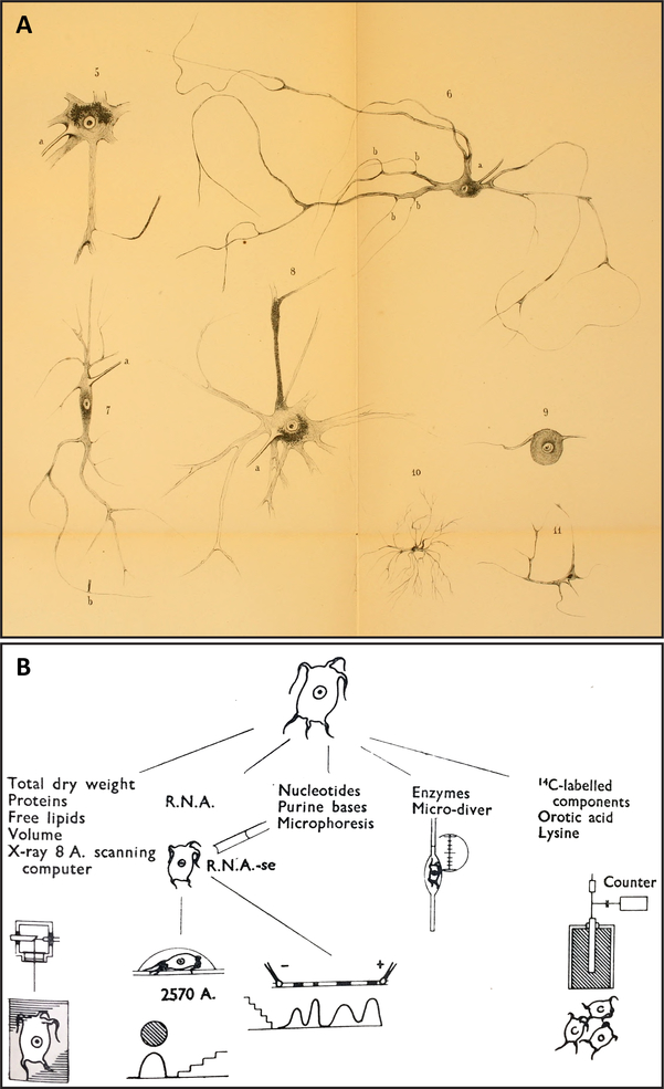
Fig. 1.
Demonstrations of single-neuron isolation procedures, nearly a century apart, which anticipate isolation methods performed at present for studying single-cell transcriptomics, proteomics, and peptidomics. (A). Plate II of Deiters (1865) [81], showing the morphologies of neurons collected from the central nervous system by hand microdissection. Note the recovery of many fine processes extended from each perikaryon, including axons and dendrites. These drawings are in the public domain. (B). Figure 2 of Hydén (1959) [187] showing a workflow schematic of possible assays that can be performed on single neurons isolated from freshly prepared tissue of the lateral vestibular nucleus. Neurons in this region are named “giant cells of Deiters” in honor of Deiters’s initial description of these cells (see [404]). Reproduced with permission from Nature Publishing Group.

