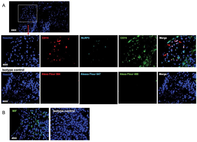
Figure 4. NLRP3 and CD74 are expressed by CD14+ cells in human acute cutaneous lupus lesion.
(A) Immunofluorescent staining of human acute cutaneous lupus lesion with antibodies to CD14 (red), NLRP3 (cyan) and CD74 (green) or control IgG. All nuclei were counterstained with Hoechst 33342. The upper panel shows nucleus staining (original magnification, ×200). The lower panels show fluorescent images for CD14, NLRP3, CD74 or control IgG staining in the areas indicated by the rectangle in the upper panels (original magnification, ×400). Arrows indicate triple-stained cells for CD14, NLRP3, and CD74. (B) Immunofluorescent staining of human acute cutaneous lupus lesion with antibodies to MIF (green) or control IgG. All nuclei were counterstained with Hoechst 33342. Representative data from 2 independent experiments.

