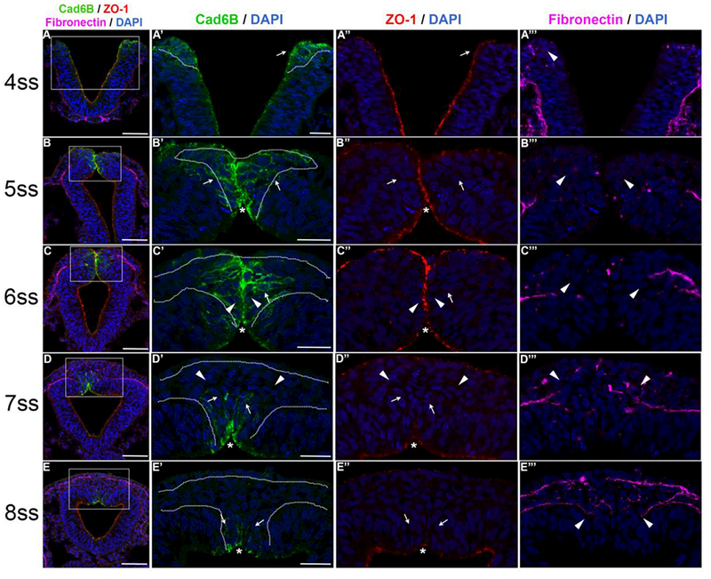
Figure 1. Cad6B levels in cranial neural crest cells undergoing EMT down-regulated dismantling of apical tight junctions and basement membrane degradation.

Representative transverse midbrain sections taken through embryos that have been immunostained for Cad6B (green), ZO-1 (red), and fibronectin (violet). DAPI stain labels cell nuclei (blue). (A-E, left column) Lower magnification images depicting triple-label immunohistochemistry (Cad6B/ZO-1/fibronectin) merged with DAPI. (A’-E’) Higher magnification images of dorsal neural tubes from section images in (A-E, highlighted by the white boxes) demonstrating Cad6B and DAPI. (A’’-E’’) The same images in (A’-E’) but showing ZO-1. (A’’’-E’’’) The same images in (A’’-E’’) but demonstrating fibronectin. All asterisks in (A’-E’) demarcate the population of Cad6B-positive cells that remain polarized and exhibit ZO-1 expression. (A-A’’’) Cad6B-positive midbrain neural crest cells at the 4ss. Epithelial premigratory neural crest cells (A’, A’’, arrows) express both Cad6B and ZO-1 at apical membranes and adhere to the basement membrane (A’’’, arrowhead). (B-B’’’) The midbrain neural crest cell domain expands at the 5ss, with most of the neural crest cells maintaining expression of Cad6B- and likely ZO-1 (B’, B’’, arrows; B’’’, arrowheads). (C-C’’’) Midbrain neural crest cells at the 6ss, when EMT is occurring en masse. Some neural crest cells at the midline have not yet initiated EMT, as they still express ZO-1 (similar to Claudin-1 at these axial levels and somite stages (Fishwick et al., 2012)) and appear polarized (O’, arrowheads). Delaminating Cad6B-positive neural crest cells (O’, C’’, arrows, and C’’’, arrowheads) appear to be oriented laterally towards the fused basement membranes. (D-D’’’) Midbrain neural crest cells at the 7ss. Leading neural crest cells exhibiting significantly diminished Cad6B levels (D’-D’’’, arrowheads) begin to emigrate through the fused basement membrane, which are followed by newly depolarized Cad6B-positive/ZO-1-negative neural crest cells (D’, D’’, arrows) that have begun to emigrate from the midline. (E-E’’’) Remaining premigratory Cad6B/ZO-1 double-positive neural crest cells (E’, E’’, arrows), and the bulk of Cad6B-negative migratory neural crest cells that have completed EMT (E’’’, arrowheads), at the 8ss. The duration between somite stages is about 1.5 hours. Scale bars in (A-E) are 50 pm. Scale bars in (A’-E’) are 20 pm and also apply to corresponding images in (A’’’-E’’’).
