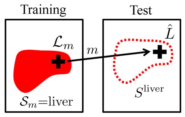
Fig. 4:
Illustration of keypoint transfer segmentation for the example of liver. The crosses indicate keypoints in the training and test images with the match m, illustrated as arrow. For the label transfer to take place, the label of the training keypoint and the voted label of the test keypoint have to be liver. The segmentation map of the training image = liver is then transferred and the probability map for liver Sliver is updated. To increase the robustness and accuracy of the segmentation, we weigh the transferred segmentation according to the certainty in (i) the keypoint label voting, (ii) the match, and (iii) the local intensity similarity of the test and training image.

