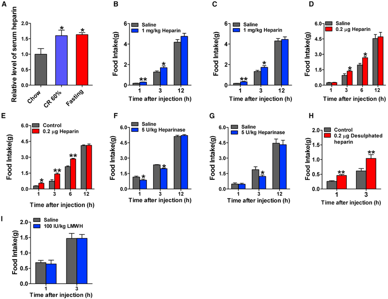
Figure 1. Heparin Synthesis at Different Nutritional States and Acute Effects of Heparin.
(A) Serum heparin levels in male C57BL6/J mice fed with normal chow, fed with CR 60% diet, or fasted for 24 hr (n = 7 per group).
(B and C) Dark-cycle food intake of male (B, n = 7 or 8 per group) and female (C, n = 7 or 8 per group) C57BL6/J mice after intraperitoneal (i.p.) injection of 1 mg/kg heparin or saline.
(D and E) Dark-cycle food intake of male (D, n = 6 per group) and female (E, n = 7 or 8 per group) C57BL6/J mice after intracerebroventricular (i.c.v.) injection of 0.2 μg heparin or saline.
(F and G) Dark-cycle food intake of male (F, n = 6 or 7 per group) and female (G, n = 6 or 7 per group) C57BL6/J mice after i.p. injection of 5 U/kg heparinase or saline.
(H) Dark-cycle food intake of male C57BL6/J mice after i.c.v. injection of 0.2 μg desulfated heparin or saline (n = 6 per group).
(I) Dark-cycle food intake of female C57BL6/J mice after i.p. injection of 100 IU/kg low-molecular-weight heparin (LMWH) or saline (n = 7 or 8 per group).
Results are presented as mean ± SEM. In (A), *p ≤ 0.05 by one-way ANOVA followed by post hoc Bonferroni tests. In (B)–(I), *p ≤ 0.05and **p ≤ 0.01 by two-way ANOVA followed by post hoc Bonferroni tests. See also Figure S1.

