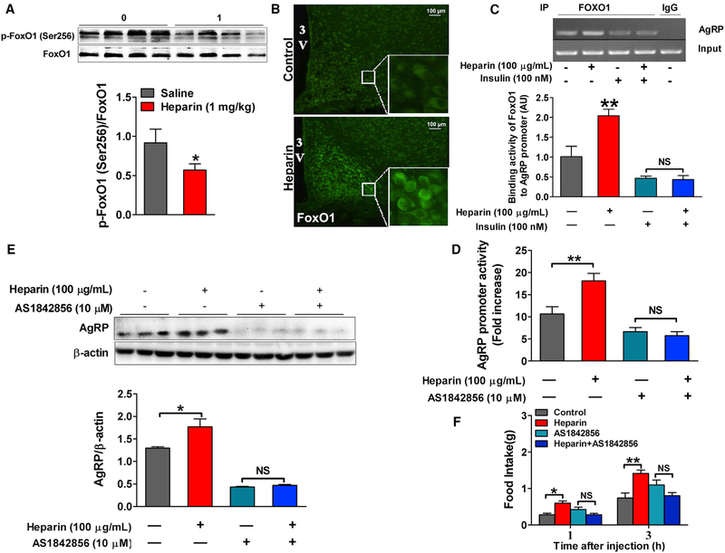
Figure 7. Heparin Promotes AgRP Expression and Secretion by FoxO1.
(A) Immunoblots and quantification of p-FoxO1/FoxO1 protein expression in the hypothalamus of male C57BL6/J mice after 16 days of saline or 1 mg/kg heparin i.p. injection (n = 8 per group).
(B) Representative images of FoxO1 immunofluorescent staining (green) in the hypothalamus of male C57BL6/J mice after 16 days of saline or 1 mg/kg heparin i.p. injection.
(C) Use of chromatin immunoprecipitation (ChIP) assays to detect the binding activity of FoxO1 to AgRP promoter in N38 cells cultured with vehicle, 100 μg/mL heparin, 100 nM insulin, or heparin + insulin (100 μg/mL+100 nM) for 3 hr (n = 6 per group).
(D) Relative luciferase activity driven by Agrp promoter (FOXO1 binding fragment) in N38 cells cultured with vehicle (control), 100 μg/mL heparin, 10 μM AS1842856 (FoxO1 antagonist), or 100 μg/mL heparin + 10 μM AS1842856 for 3 hr (n = 6 per group).
(E) Immunoblots and quantification of AgRP protein expression in N38 cells cultured with vehicle, 100 μg/mL heparin, 10 μM AS1842856, or 100 μg/mL heparin + 10 μM AS1842856 for 12 hr (n = 6 per group).
(F) Dark-cycle food intake of female C57BL6/J mice after i.c.v. injection of saline, 0.2 μg heparin, 20 pmol AS1842856, or heparin + AS1842856 (0.2 μg + 20 pmol) (n = 6 per group).
Results are presented as mean ± SEM. *p ≤ 0.05 and **p ≤ 0.01 by two-way ANOVA followed by post hoc Bonferroni tests. See also Figure S7.

