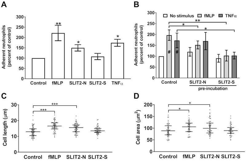
Figure 4. Slit2-N and Slit2-S have different effects on cell adhesion and cell area.
(A) Neutrophils were allowed to adhere to fibronectin for 30 minutes, and Slit2 proteins, fMLP, TNFα, or an equal volume of buffer (control) were added for an additional 30 minutes. Plates were then washed, and adherent cells were air-dried, stained, and counted. Values are mean ± SEM, n = 4. (B) Cells were pre-incubated for 15 minutes with either Slit2-N or Slit2-S before the addition of fMLP or TNFα for an additional 15 minutes. Cells were then allowed to adhere to fibronectin for 30 minutes. Adherent cells were air-dried, stained, and counted. Values are mean ± SEM, n = 4. # indicates p < 0.05 compared to the no stimulus control (t test). (C-D) Neutrophils were incubated with a point source of buffer, Slit2 proteins, or fMLP for 10 minutes, then fixed and (C) cell length and (D) cell area were measured with Image J. The results are mean ± interquartile range of 20 cells analyzed from three different donors. * indicates p < 0.05, *** p < 0.001 (1-way ANOVA, Dunnett’s test).

