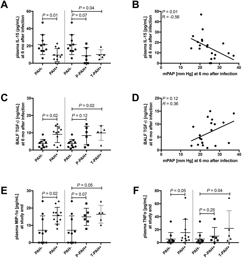
Figure 8.
Compared with PAH– animals, macaques developing PAH+ showed higher baseline plasma levels of chemokines involved in monocyte–macrophage trafficking and elevated levels of growth factor TGFβ in BALF at 6 mo after infection. We quantified a total of 31 cytokines, chemokines, and growth factors in serial plasma and BALF samples. Differences in inflammatory markers between PAH+ and PAH–macaques at 6 mo after infection and at study end were assessed by using Mann–Whitney tests. To test for associations between mPAP and each immune marker, Spearman correlation was used. All graphs of correlation analysis, show regression lines.(A) At 6 mo after infection, plasma levels of IL15 were decreased (P = 0.01) in PAH+ compared with PAH– macaques, and (B) IL15 and mPAP were negatively correlated (P = 0.01). In BALF, (C) TGFβ levels were increased (P = 0.02; Figure 8 C) in PAH+ compared with PAH– animals and (D) trended toward a significant correlation with mPAP (P = 0.12). At study termination, plasma levels of (E) MIPα (P = 0.02) and (F) TNFα were increased (P = 0.2 and P = 0.05, respectively) in PAH+ compared with PAH– animals.

