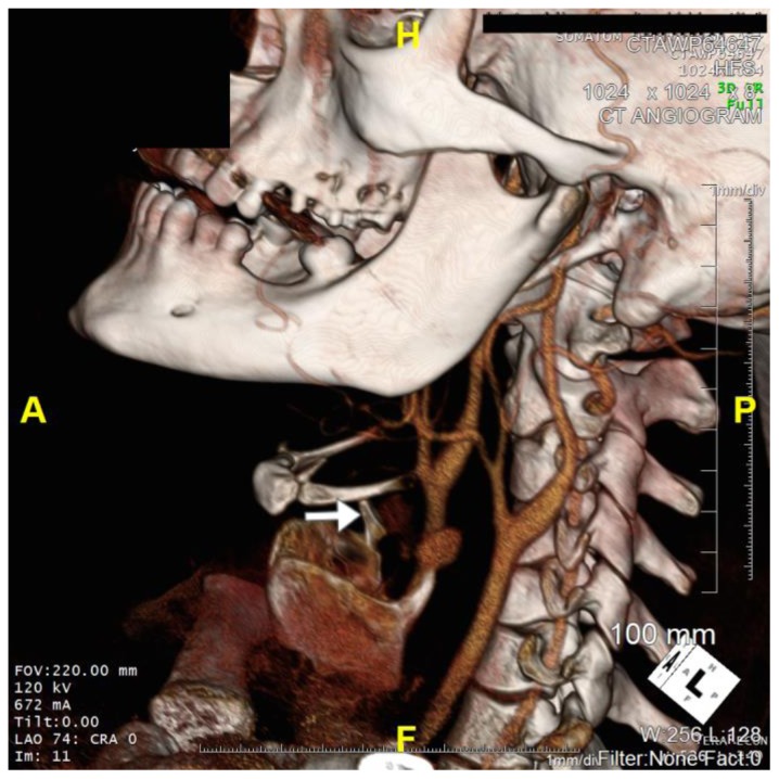
Figure 5.
36 year old male imaged following blunt force trauma to right side of the neck resulting in a right carotid artery pseudoaneurysm.
Findings: Fracture of the right superior horn of the thyroid cartilage (white arrow), with lateral bowing of the common carotid artery proximal to the carotid bifurcation suggesting extramural compression from the pseudoaneurysm seen on CTA imaging
Technique: Siemens SOMATOM 64 slice CT scanner, Axial Left Anterooblique (LAO) projection 3D CT reformat, 672mA, 120kV, 1mm slice thickness, Tilt 0 degrees, LAO 74 degrees, Cranial 0 degrees)

