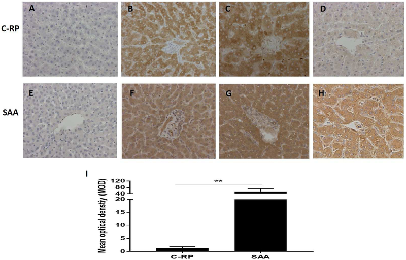
Figure 3: Expression of C-RP (above) and amyloid A (below) in liver tissue detected by IHC (magnification x200).
(A, E) Expression of C-RP and amyloid A in a naive pig liver (negative control); (B, F) Expression of C-RP and amyloid A in the liver of a baboon that experienced anaphylactic shock (without transplantation) (positive control). (C, G) Expression of C-RP and amyloid A in the liver of a baboon with a pig kidney graft that underwent hyperacute rejection (while receiving the IL-6R inhibitor, tocilizumab, and the TNF-α antagonist, etanercept) (positive control). (D, H) Expression of C-RP and amyloid A in the liver of a representative baboon with a pig kidney graft that was undergoing antibody-mediated rejection (while receiving tocilizumab). There is considerable expression of amyloid A, but not C-RP, in the liver. (I) Comparison of C-RP and amyloid A expression in the livers of baboons with pig kidney grafts (while receiving tocilizumab). The difference in expression is significant (**p<0.01).

