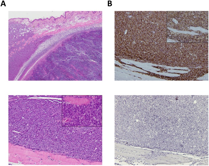
Fig 3. Histopathological characteristics of the CLBL-1GFP+Luciferase+ cell line as a xenograft tumor in SOPF/SHO SCID mice.
Mouse interscapular region. Xenograft CLBL-1GFP+Luciferase+ tumor. (A) Upper panel—Compact infiltration of the dermis, hypodermis, muscle panniculus and skeletal muscle by large lymphoid cells (H&E, 20x). Bottom panel–Magnification of the tumor showing lymphoid cells with indistinct cytoplasmic borders and finely distributed nuclear chromatin and inconspicuous nucleolus. A muscle fiber is surrounded by tumor cells in the insert (H&E, 100x, insert 400x). (B) Upper panel–Immunohistochemistry for B-cells showing positivity in virtually 100% of the tumor cells (anti-CD20 antibody, Gill’s hematoxylin, 100x, insert 400x). Bottom panel–Immunohistochemistry for T-cells, showing that tumor cells were negative for this marker (anti-CD3, Gill’s hematoxylin, 100x).

