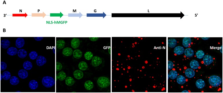
Fig 1. Recombinant RABV ERA-NLS-hMGFP.
(A) The schematic representation of recombinant RABV ERA genome with the inserted NLS-hMGFP. 3’ and 5’ denotes the direction of negative sense genome. N, P, M, G and L corresponds to nucleoprotein, phosphoprotein, matrix protein, glycoprotein and large RNA dependent RNA polymerase genes, respectively. (B) BSR cells were infected with recombinant RABV ERA-NLS-hMGFP virus for 24 h at 37°C. The cells were stained with DAPI (blue) and anti-N monoclonal antibodies (red) and imaged. The merge represents the co-localization of NLS-hMGFP with DAPI demonstrating nuclear targeting of GFP (green).

