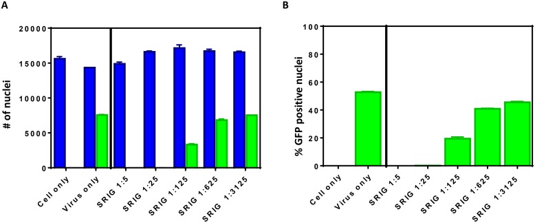
Fig 2. Quantification of DAPI and GFP positive cells by HTNT.
BSR cells were infected with ERA-NLS-GFP for 20 h at 37°C in the presence and absence of SRIG and stained with DAPI (for nucleus). The number of DAPI and GFP positive nuclei are quantitated. (A) Total number of DAPI stained nuclei (corresponding to number of cells) and GFP co-localization with DAPI stained cells are shown as blue and green bars, respectively. (B) The percent GFP positive cells under various conditions were calculated.

