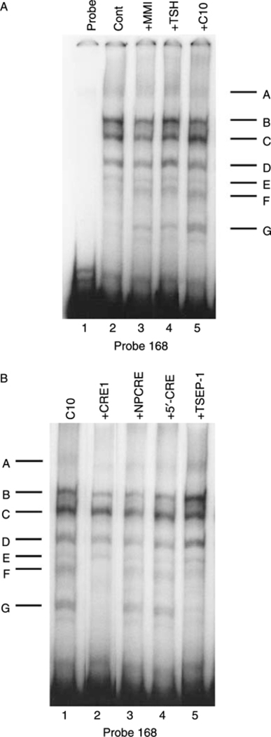
Figure 4.

Effects of MMI and C10 on binding of proteins to the −168 bp probe. Representative EMSAs performed using cell extracts, as detailed in Materials and Methods, and the −168 bp probe, which spans the region between −168 and +1 bp from the transcription start site of the PD1 gene. (A) FRTL-5 cells were grown and prepared as detailed in Materials and Methods, with final treatments in 5H medium as indicated with 0·5% DMSO (Control), 5 mM MMI, 1×10⁻¹⁰ M TSH, or 0·5 mM C10 for 36 h. Protein-DNA complexes are labeled as A-G (see text). Lane 1, probe alone. (B) FRTL-5 cells were grown and prepared as detailed in Materials and Methods, with the probe incubated in 5H medium with 0·5 mM C10 alone or with 100-fold excess of unlabeled competitors as indicated for 36 h. Protein-DNA complexes are labeled as A-G (see text). Similar results were obtained using cell extracts from cells treated with 5 mM MMI (data not shown).
