Figure 2.

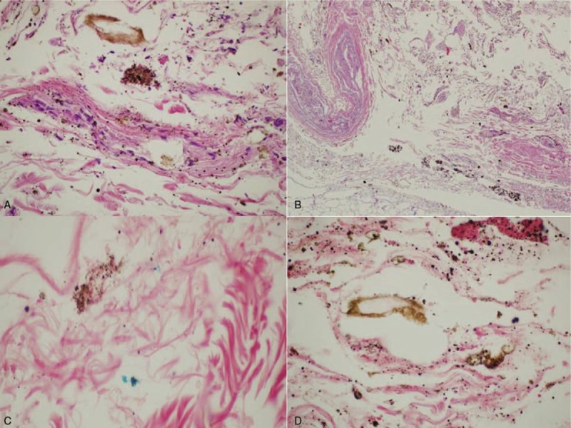
(A, B) Neutrophil infiltration of the esophageal mucosa and submucosa (H&E 40×), (H&E 4×). (C, D) Digested material and the presence of hemosiderin accumulations in the esophageal mucosa and submucosa (Perls 60×).

(A, B) Neutrophil infiltration of the esophageal mucosa and submucosa (H&E 40×), (H&E 4×). (C, D) Digested material and the presence of hemosiderin accumulations in the esophageal mucosa and submucosa (Perls 60×).