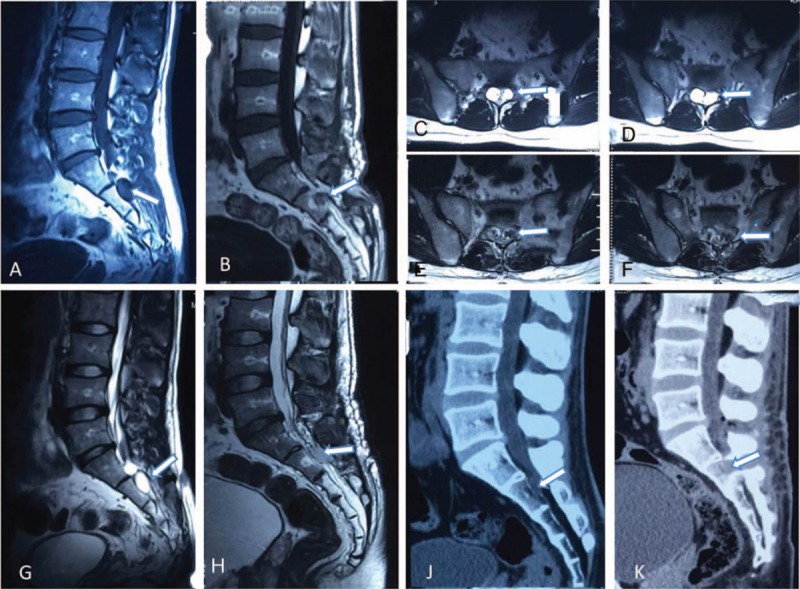
Figure 1.

A, Sagittal MRI T1-weighted image of a sacral canal cyst (arrow). B, Postoperative T1-weighted image showing sacral canal cyst disappearance (arrow). C and D, Axial MRI T2-weighted images showing 2 sacral canal cysts (arrows). E and F, Postoperative axial MRI T2-weighted images showing sacral canal cyst disappearance (arrows). G, Sagittal MRI T2-weighted image showing multiple sacral canal cysts (arrow). H, Postoperative T2-weighted image showing the disappearance of the sacral canal cysts (arrow). J, Sagittal CT reconstruction image of the sacral canal cyst (arrow). K, Postoperative sagittal CT reconstruction image showing sacral canal cyst disappearance (arrow) and lamina restoration. CT = computed tomography, MRI = magnetic resonance imaging.
