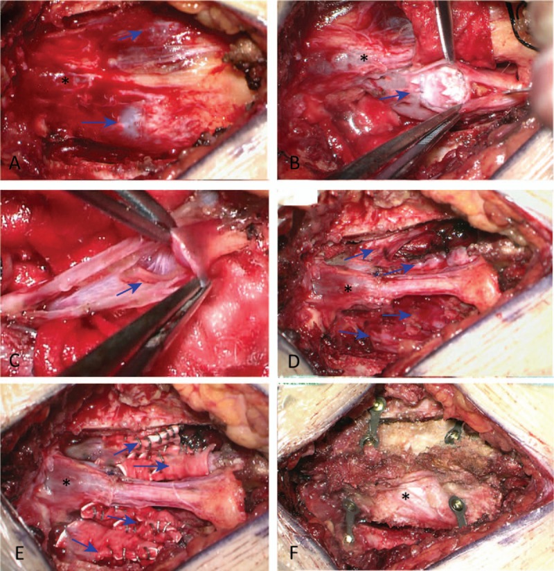
Figure 3.

A, End of the dural sac in the sacral canal after lamina removal (∗), with multiple sacral canal cysts in the tail end (arrow). B, Bone occupying lesions in the cyst (arrow). C, Nerve root which tortuously travels and adheres to the cyst wall (arrow). D, Nerve root sleeve plasty after partial removal of the cyst wall (arrow) (∗, end of the dural sac). E, Post-radiculoplasty by artificial dura mater wrapping (arrow) (∗, end of the dural sac). F, ∗Restored lamina fixed with titanium nails and plates.
