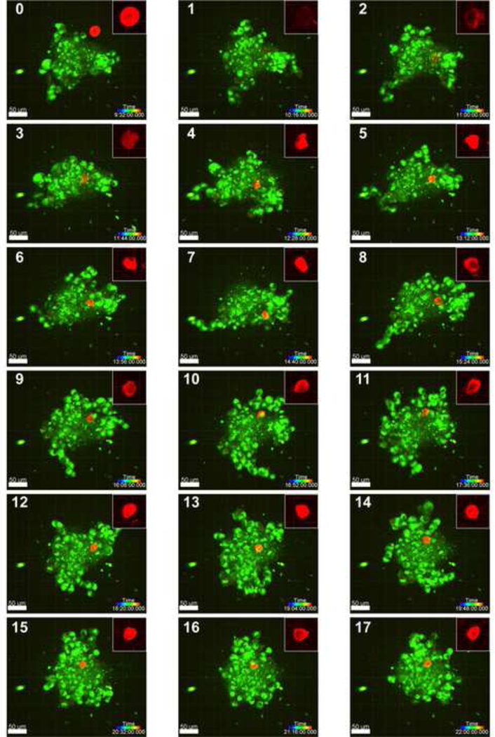
Figure 4.

Error! No text of specified style in document.. Confocal 3D projections of a representative soft MP incorporating into a 7-day old A375 spheroid, with images taken at 44- minute intervals (MP: red, cells: green; scale bar = 50 μm; rainbow bar = time progression). The soft MP first interacts with and integrates into the spheroid between hour 9 and 10 of imaging (panels 0 and 1) and continues to be moved within the spheroid through hour 22. Insets (50 μm x 50 μm) depict the substantial deformation of soft MPs. Images with the MP outside and not interacting with the spheroid (< 9.5 hours, panel 0) were not included in the figure.
