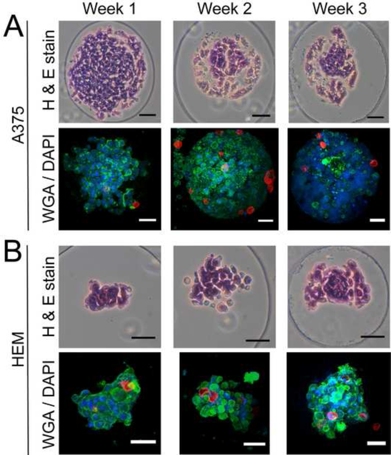
Figure 5.

Histology (cell-only spheroids) and confocal imaging (spheroids with soft MPs) of HEM and A375 spheroids over 3 weeks. (A) A375 spheroids successfully incorporated soft MPs after a 1-week maturation period. At the 2- and 3-week time points, A375 sphero ids exhibited features associated with cell death possibly induced by the continued proliferation and crowding that could limit nutrient diffusion into the centroid of the spheroids. (B) HEM spheroids that were matured for up to 3 weeks prior to adding MPs exhibited matrix and melanin deposition (black dots). MPs added to the HEM spheroids after 1, 2, and 3-week maturation periods successfully incorporated. (Scale bar = 50 μm).
