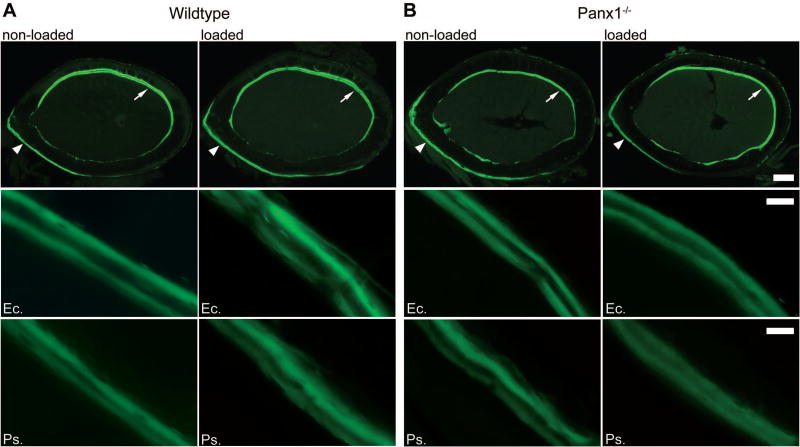
Figure 3.
Load-induced changes in the periosteal and endocortical surfaces of femoral midshaft after 2 weeks of loading in wildtype and Panx1−/− mice. Top panels: Representative cross-section images showing double calcein labels for age-matched non-loaded and loaded wildtype (A) and Panx1−/− (B) bones. Scale bar = 200 µm. Middle panels: Representative images of double calcein labeling on lateral endocortical (Ec.) surfaces (indicated with arrows in the top panels) of non-loaded and loaded wildtype and Panx1−/− bones. Scale bar = 20 µm. Bottom panels: Representative images of double calcein labeling on medial periosteal (Ps.) surfaces (indicated with arrowheads in the top panels) of non-loaded and loaded wildtype and Panx1−/− bones. Scale bar = 20 µm.

