. 2018 Dec 26;21(4):453–462. doi: 10.4048/jbc.2018.21.e62

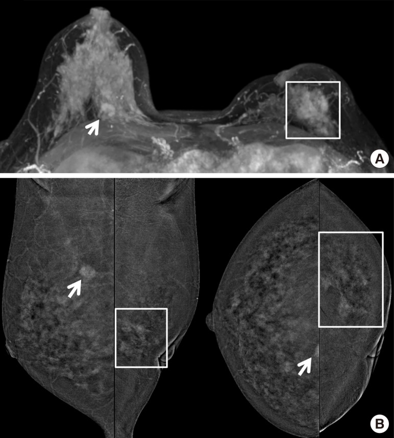
Figure 3. An asymptomatic 49-year-old woman confirmed with ductal carcinoma in situ (DCIS) on her left breast with previous history of breast-conserving surgery due to invasive ductal carcinoma on ipsilateral side of breast. (A) On contrast-enhanced magnetic resonance imaging (CEMRI), symmetric severe background parenchymal enhancement was noted. Axial maximal intensity projection image showed index cancer with regional non-mass enhancement (square) around previous operation site on her left breast and unexpected enhancing mass (arrow) in contralateral right breast. (B) On contrast-enhanced digital mammography, there was a regional non-mass enhancement correlated with index cancer (square) in left lower outer quadrant. And contralateral irregular enhancing mass (arrow) was also seen in right upper medial breast, identical to CEMRI. She underwent bilateral mastectomy, and the result was DCIS for left breast, and invasive tubular carcinoma for contralateral right breast (true positive).

Figure 3