. 2018 Dec 26;21(4):453–462. doi: 10.4048/jbc.2018.21.e62

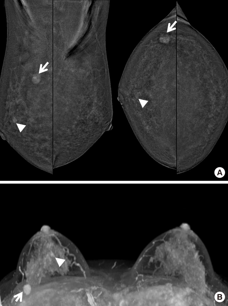
Figure 4. A 38-year-old woman with proven invasive ductal carcinoma in her right breast. (A) She underwent contrast-enhanced magnetic resonance imaging, and axial maximal intensity projection image showed index cancer (arrows) with unexpected subareolar enhancing small mass (arrowheads) in right breast. (B) On contrast-enhanced digital mammography, the index cancer (arrow) was well visualized with another subareolar enhancing mass (arrowhead) in right breast. She underwent breast-conserving surgery for right breast, and the result was single invasive ductal carcinoma. A subareolar small enhancing mass was false positive lesion which confirmed as fibroadenomatoid lesion.

Figure 4