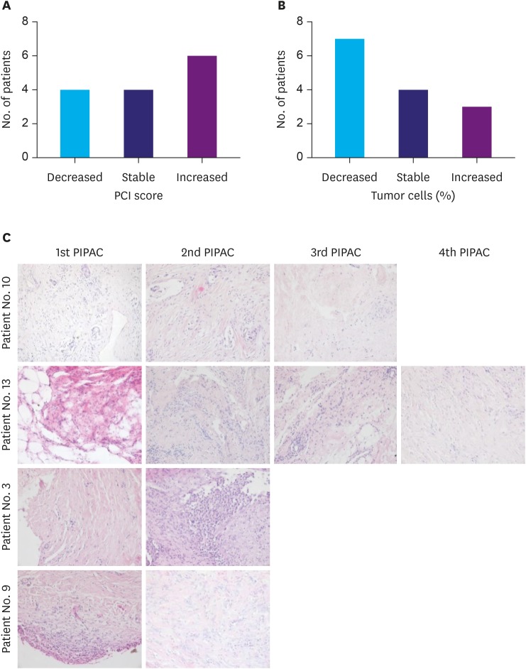
Fig. 2. PCI and tumor cell portion developments in patients with multiple PIPAC procedures. (A) Number of patients with decreased, stable, and increased PCI, (B) Number of patients with decreased, stable, and increased tumor cell portions in PM biopsies, and (C) representative H&E staining of patients with decreased tumor cell portions (patients No. 10 and No. 13) and increased tumor cell portions (patients No. 3 and No. 9). The biopsies for tumor cell evaluation were taken before each PIPAC.
PCI = peritoneal carcinomatosis index; PIPAC = pressurized intraperitoneal aerosol chemotherapy; PM = peritoneal metastasis; H&E = hematoxylin and eosin stain.

