Fig. 5.
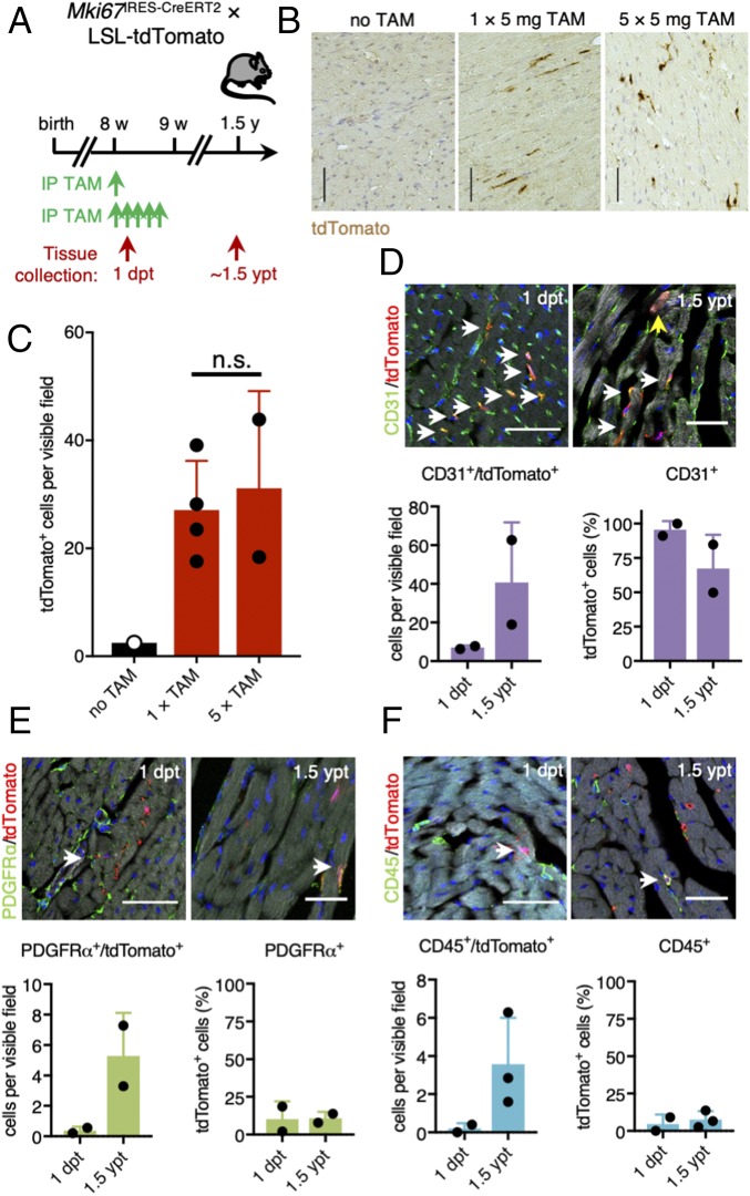
Continuous cellular turnover of noncardiomyocyte lineages during adult homoeostasis of the murine heart. (A) Timeline of tamoxifen injection and tissue collection in adult Mki67IRES-CreERT2 × LSL-tdTomato mice. (B and C) Representative images of stained paraffin sections (B) and corresponding quantification (C) of tdTomato labeling 1.5 y after tamoxifen exposure (1.5 ypt). Mice were either injected once with 5 mg of tamoxifen (n = 4 mice) or five times with 5 mg of tamoxifen (n = 2 mice). Noninjected mice were used as control (n = 1 mouse). (Scale bars: 100 μm.) (D–F) Representative images and quantification of costainings of tdTomato-traced cells (red) at 1 dpt and 1.5 ypt and CD31+ (D), PDGFRα+ (E), or CD45+ cells (F) (green) (n = 2–3 mice). ypt, years post tamoxifen. White arrows point at double-positive cells. The yellow arrow shows one of the tdTomato-labeled cardiomyocytes we found across sections. Nuclei were counterstained with DAPI (blue). Phalloidin (gray) was used to stain polymerized F-actin. [Scale bars: 50 μm (Upper) and 30 μm (Lower).]

