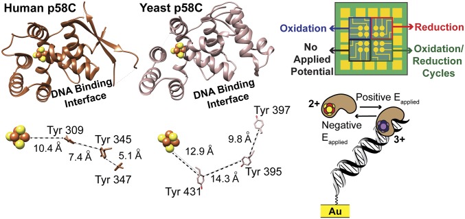
Fig. 1.
Yeast and human p58C structures can both support a redox switch. (Left) Comparison of p58C structures (Upper) and conserved tyrosines (Lower) near the [4Fe4S] cluster in yeast p58C (light brown) and human p58C (dark brown). Structures rendered using human p58C [Protein Data Bank (PDB) ID code 3L9Q] (15) and yeast p58C (PDB ID code 6DI6, SI Appendix, Table S3) structures. (Right) Diagram of the multiplexed DNA electrochemistry platform (Upper) and a cartoon depicting the change in DNA binding associated with redox switching (Lower). Human p58C image adapted from ref. 15. Multiplex chip and yeast p58C images from ref. 9. Reprinted with permission from AAAS.

