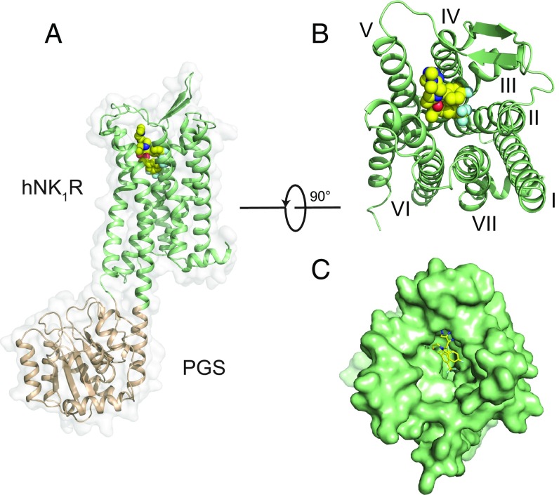
Fig. 1.
Structural features of hNK1R; (A) Global structure of the hNK1R-PGS fusion protein from side view. The hNK1R is represented as a green cartoon, with the PGS domain (wheat) fused between TMs 5 and 6. L760735 is shown as spheres with yellow carbons. (B) Extracellular view of hNK1R, with PGS domain removed. (C) Surface representation as in B.

