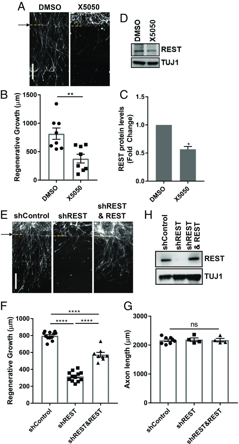
Fig. 6.
REST expression is required for axon regeneration. (A) DRG spot-cultured neurons were treated with DMSO or 50 μM X5050 10 min before injury, fixed, and stained for SCG10 40 h after axotomy. Arrow and dotted lines indicate the axotomy site. (B) Regenerated axon length of DRG spot-cultured neurons was measured 40 h after axotomy from images in A (Individual data plotted; n = 8 biological replicates; **P < 0.01 by t test; mean ± SEM). (C) Normalized intensity was calculated from Western blot analysis of DRG spot-cultured neurons after treatment of DMSO or 50 μM X5050 (n = 3 biological replicates, *P < 0.05 by t test; mean ± SEM). (D) Representative image from Western blot analysis of DRG spot-cultured neurons treated with DMSO or 50 μM X5050 for 24 h. Expression levels of REST were analyzed by Western blot. TUJ1 serves as a loading control. (E) DRG spot-cultured neurons were infected with lentivirus expressing control shRNA (shcontrol), shRNA targeting REST or human REST, fixed, and stained for SCG10 40 h after axotomy. Arrow and dotted lines indicate the axotomy site. (F) Regenerative axon growth was measured 40 h after axotomy from images in E. Individual data plotted; n = 8–12 biological replicates; ****P < 0.0001 by one-way ANOVA; mean ± SEM. (G) DRG spot-cultured neurons were infected with the indicated lentivirus, fixed, and stained for TUJ1 at DIV7 (n = 4–8 biological replicates for each; ns, not significant by one-way ANOVA; mean ± SEM). (H) DRG spot-cultured neurons were infected with control shRNA (shcontrol), shRNA-targeting REST, or human REST lentivirus. Expression levels of REST was analyzed by Western blot. TUJ1 serves as a loading control. (Scale bars: 200 μm.)

