Fig. 1.
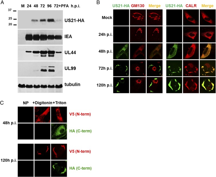
Characterization of US21 protein expression. (A) Kinetics of pUS21 expression during HCMV infection. HFFs were infected with TRUS21HA [multiplicity of infection (MOI) of 1 pfu/cell]. At the indicated times p.i., protein cell extracts were analyzed by immunoblotting with anti-HA, IEA, UL44, UL99, or tubulin MAbs. The expression levels of IEA (IE1 and IE2), UL44, and UL99 were assessed as controls for representative IE, E, and L proteins, respectively. Cell extracts were from mock-infected cells (M); cells infected for 24, 48, 72, and 96 h; or cells infected and treated with phosphonoformic acid (PFA) (200 μg/mL) for 72 h. (B) Intracellular localization of pUS21HA during HCMV replication cycle. HFFs were infected with TRUS21HA (MOI of 1 pfu/cell) or mock infected, and, at various times p.i., cells were fixed, permeabilized, and immunostained with an anti-HA (green) and either (Left) GM130 (Golgi marker, red) or (Right) CALR (ER marker, red) MAbs. (C) Membrane topology of pUS21. HFFs were infected with TRUS21NV5-CHA (MOI of 1 pfu/cell), and, at 48 h or 120 h p.i., cells were not permeabilized (NP), or selectively or completely permeabilized using digitonin (+Digitonin) or Triton X-100 (+Triton). The pUS21 N- and C-terminal tags were then immunostained with MAbs to V5 and HA, respectively. Images in B and C are representative of three independent experiments. (Magnification in B and C: 60x.)

