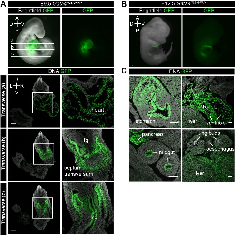
Fig. 6.
Gata4H2B-GFP/+ reporter expression at midgestation. (A) Upper panel showing lateral whole-mount immunofluorescence of Gata4H2B-GFP/+ embryos at E9.5. GFP is overlaid over bright field or GFP alone. (a-c) Transverse sections of an E9.5 Gata4H2B-GFP/+ embryo at indicated positions on whole-mount embryo. GFP was visualised directly and DNA was stained with Hoechst (a) GFP continues to be expressed throughout the heart (b) and the septum transversum. (c) GFP is also expressed in the midgut (mg) epithelium at this stage but is absent from the foregut and hindgut. (B) Lateral whole-mount immunofluorescence of Gata4H2B-GFP/+ embryos at E12.5. GFP is overlaid over bright field or GFP alone. (C) Transverse sections of an E12.5 Gata4H2B-GFP/+ embryo. GFP was visualised directly and DNA was stained with Hoechst. GFP expressing cells are present in the stomach epithelium and associated mesenchyme, the liver mesenchyme, the endocardium and myocardium of the heart, pancreas, midgut epithelium and the mesenchyme but not epithelium of the lung buds. Scale bars: 100 μm. Neural tube (NT), foregut (fg), hindgut (hg), right (R), left (L), dorsal (D), ventral (V), anterior (A), posterior (P).

