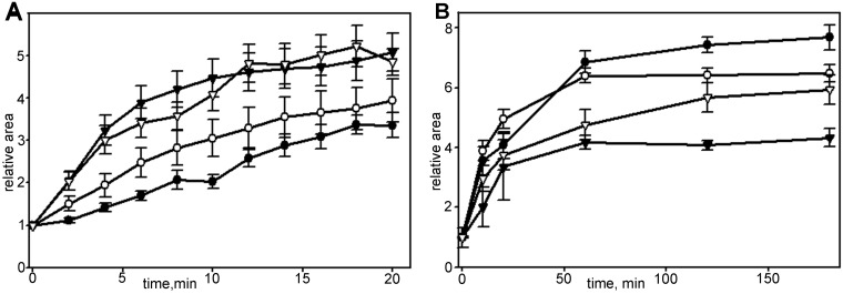
Fig. 6.
Changes in spreading kinetics of Vero cells in the presence of cytochalasin D. For each set N=20, data presented as mean±s.e.m. (A) Spreading in first 20 min in control (black triangles), blebbistatin-treated (white triangles), cytochalasin D-treated (black circles) and cells under simultaneous addition of cytochalasin D and blebbistatin (white circles). (B) Spreading in first 180 min in control (black circles), treated with blebbistatin (white circles), treated with cytochalasin D (black triangles) and cells under simultaneous treatment with cytochalasin D and blebbistatin (white triangles). Myosin relaxation facilitates overall spreading process.

