Fig. 8.
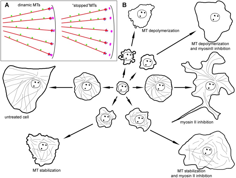
Schematic representation of the role of dynamic MTs and myosin relaxation in cell spreading. (A) MTs (red arrows) are responsible for the delivery of signals that are transported along MTs towards the cell edge (green circles), while other factors are delivered directly by growing MT tips along with plus end comet (blue circles). Nascent focal adhesions are in purple. MTs stopped by nocodazole and taxol treatment do not reach focal adhesions with their plus-end comets. (B) Untreated cell forms a large lamellum during fast spreading module and transfers to polarization during slow spreading. A cell with depolymerized MTs loses fast spreading module and demonstrate continuous blebbing. When MTs are stabilized, kinetics remains the same as for depolymerized MTs, but blebbing almost disappears. The inhibition of myosin II does not affect the fast spreading module, but heavily alters cell morphology during slow spreading module. Additional treatment of cell with compromised MTs with myosin II pathway inhibitors partially restores kinetics of fast spreading.

