Fig. 2.

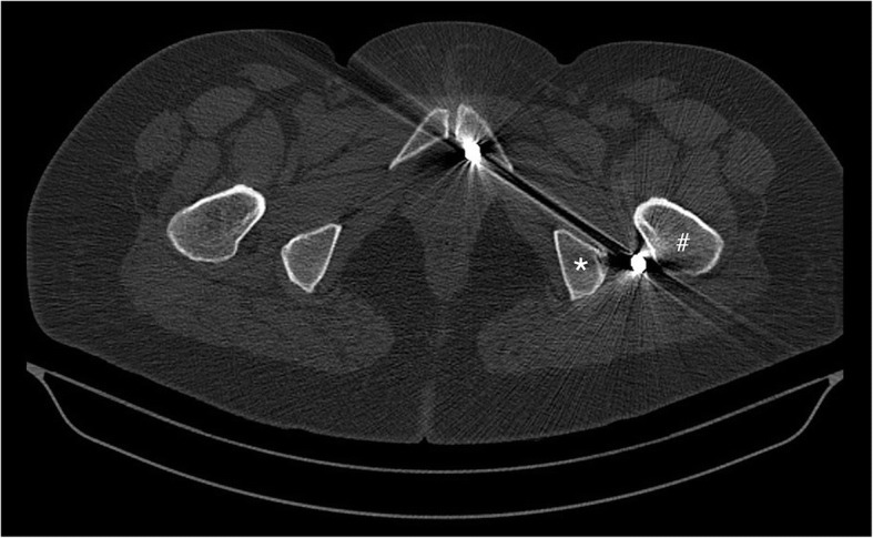
Preoperative computed tomography scan demonstrating the pellet of interest located between the trochanter major (#) and ischial tuberosity (*) close to the course of sciatic nerve

Preoperative computed tomography scan demonstrating the pellet of interest located between the trochanter major (#) and ischial tuberosity (*) close to the course of sciatic nerve