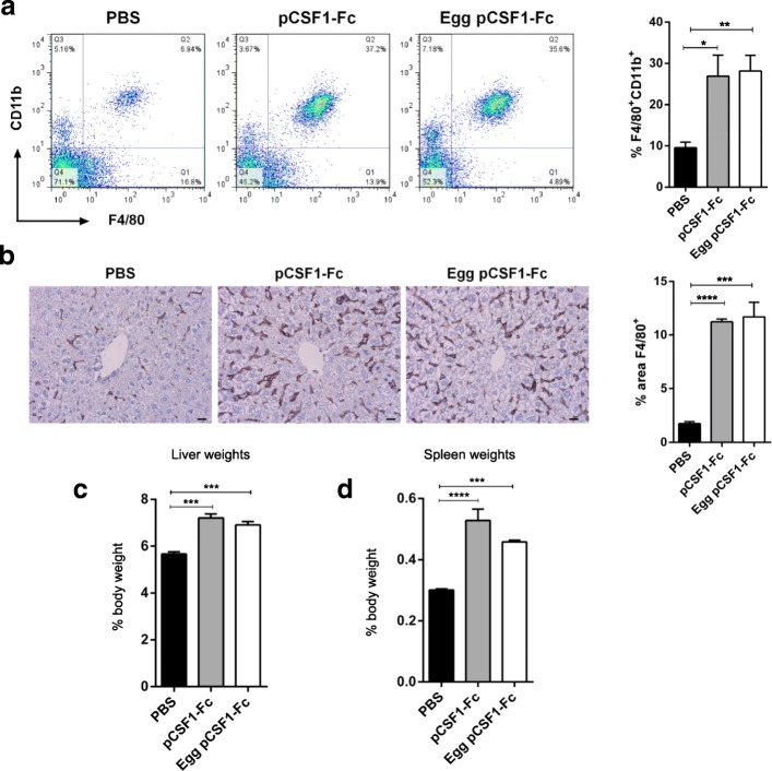
Fig. 3.
The effect of egg purified pCSF1-Fc treatment in mice is identical to that of pCSF1-Fc from CHO cells. Mice were injected with PBS, 1 μg/g pCSF1-Fc or 1 μg/g egg purified pCSF1-Fc for 4 days prior to sacrifice on day 5. a EDTA-blood was collected via cardiac bleeds and the percentage of F4/80+CD11b+ myeloid cells were determined by flow cytometry. Graph shows the mean + SEM. Significance is indicated by *p = 0.0163 and **p = 0.0037 using a t-test; n = 4. b Formalin-fixed paraffin-embedded livers were stained with an antibody against F4/80. The percentage of F4/80 staining was determined using ImageJ. Graph shows the mean + SEM. Significance is indicated by ***p = 0.0004 and ****p < 0.0001 using a test-test; n = 4. c Livers were weighed and percent body weight calculated. Graphs show the mean + SEM. Significance is indicated by ***p = 0.0004 using a t-test. There was no significant difference between the two pCSF1-Fc populations (p = 0.2482). d Spleens were weighed and percent body weight calculated. Graphs show the mean + SEM. Significance is indicated by ****p < 0.0001 and ***p = 0.0009 using a t-test. There was no significant difference between the two pCSF1-Fc populations (p = 0.1122)

