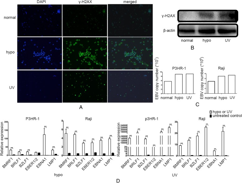
Fig. 4.
DNA damage reactivates EBV. a Expression of γ-H2AX as measured by immunofluorescence after treatment with hypoxia or UV. EBV-positive Raji cells were stained with anti-γ-H2AX and DAPI. Expression of γ-H2AX is indicated as green loci. DAPI was used to stain the cell nuclei. The merge images present the DAPI and FITC as blue and green, respectively. b Expression of γ-H2AX as measured by western blotting after treatment with hypoxia or UV in Raji cells. β-actin served as an loading control. c EBV copy number as measured by real-time PCR after treatment with hypoxia or UV. d Relative expression of EBV genes as measured by real-time PCR after treatment with hypoxia or UV

