
Figure 1:
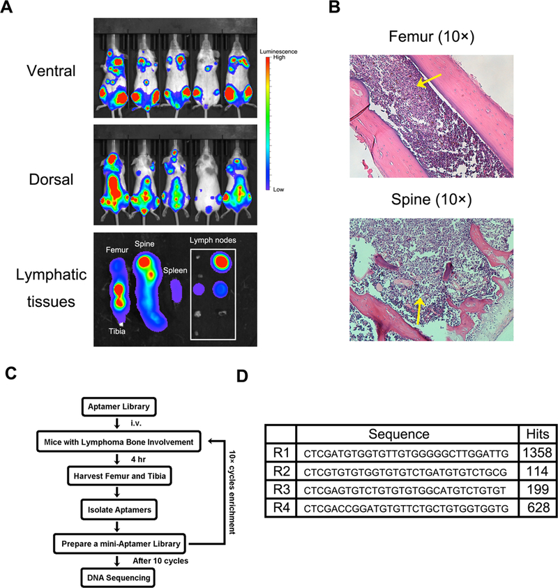
Murine model of human Burkitt’s lymphoma with bone marrow involvement for in vivo SELEX of aptamer. (A). Growth of Raji cells with luciferase gene in SCID/Beige mice was monitored by an Xenogen IVIS-200 system. Pictures were taken on both ventral and dorsal sides. The ex vivo images showed lymphoma in lymphatic tissues (femur, tibia, spine, spleen and lymph nodes). Lymphoma cells widely invaded into bones and lymph nodes, but less were observed in spleen. (B). Histological analysis of Raji lymphoma in medullary cavities of spine and femur. Raji cells invaded into bone marrow and effaced normal tissues. Yellow arrows indicate the tumor nodules. (C). Schematic diagram of in vivo SELEX process.