Figure 5.

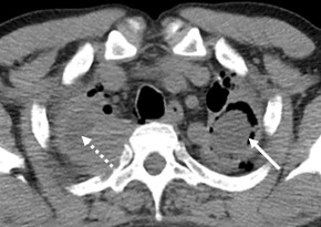
Left upper lobe cavity lesion with solid content (continuous line); Right apical thickening of the pleurae, with highlighting interior fluid (dotted line)

Left upper lobe cavity lesion with solid content (continuous line); Right apical thickening of the pleurae, with highlighting interior fluid (dotted line)